
A retinal detachment needs prompt treatment. When the retina pulls away from the back of the eye, it can no longer function normally, and vision can be affected.
Depending on the location and extent of the detachment, pneumatic retinopexy for retinal detachment may be one treatment option. It uses a small gas bubble to help move the retina back into position while laser or cryotherapy seals the retinal tear.
What Is Pneumatic Retinopexy?
Pneumatic retinopexy is a procedure used to repair certain retinal detachments without making a large surgical incision.
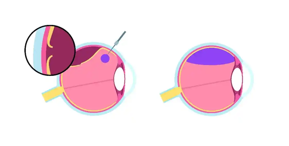
A retina specialist places a small gas bubble inside the eye. The bubble rises and presses against the detached part of the retina, helping move it back toward the eye wall.
The retinal tear is then treated with laser or cryotherapy. As the treated area heals, scar tissue forms around the tear and helps keep the retina in place.
This approach is not suitable for every retinal detachment. The location of the tear, the extent of the detachment, and other retinal findings all matter when choosing treatment.
How Does Pneumatic Retinopexy for Retinal Detachment Work?
The treatment has two main goals: reposition the retina and seal the retinal tear.
During pneumatic retinopexy for retinal detachment:
- The eye is numbed. Local anesthesia is used before treatment.
- A gas bubble is placed inside the eye. The bubble is positioned so that it can support the detached area.
- he retina is repositioned. Head positioning allows the bubble to press against the retinal tear and detached area.
- The tear is sealed. Laser or cryotherapy is applied around the tear to create scar tissue.
- The bubble gradually disappears. The eye naturally absorbs the gas over time.
Following the recommended head position is an important part of recovery because the bubble needs to remain against the treated area while the retina heals.
Who May Need Pneumatic Retinopexy?
Pneumatic retinopexy is generally considered for selected retinal detachments. It may be appropriate when the retinal tear and detached area can be effectively supported by the gas bubble.
A retina specialist will examine the retina carefully before recommending the procedure.
Other retinal detachment surgery options include:
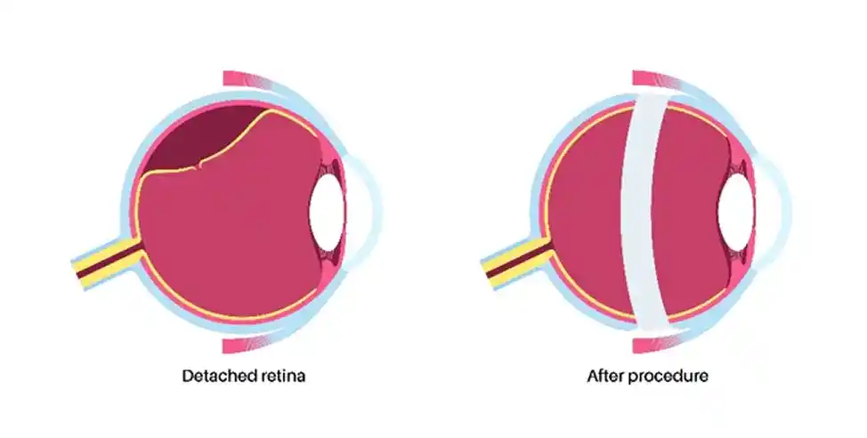
- Vitrectomy: Removes the vitreous gel so the surgeon can repair the retinal tear and reattach the retina.
- Scleral buckle: Places a supportive band around the outside of the eye to help relieve traction on the retina.
- Pneumatic retinopexy: Uses a gas bubble to reposition the retina and seal the retinal break.
In some cases, treatments may be combined.
What Happens During a Pneumatic Retinopexy Procedure?
The pneumatic retinopexy procedure is usually performed on an outpatient basis.
After numbing the eye, the retina specialist injects a small gas bubble into the eye. The bubble rises naturally, and positioning the head correctly allows it to press against the retinal tear.
The retinal break is then treated with laser or cryotherapy.
The procedure itself is relatively brief, but treatment does not end when you leave the office. Positioning, follow-up visits, and monitoring are important parts of recovery.
How Long Does Pneumatic Retinopexy Take?
How long does pneumatic retinopexy take? The procedure itself is usually short, although the exact time depends on the retinal detachment and the treatment required.
The gas bubble remains in the eye longer than the procedure itself. It gradually becomes smaller as the body absorbs it.
Your retina specialist will explain how long the bubble is expected to remain and how long you may need to follow positioning or activity restrictions.
Is Pneumatic Retinopexy Painful?
Is pneumatic retinopexy painful? The eye is numbed before the procedure, so significant pain during treatment is generally not expected.
Some patients may feel pressure or mild discomfort during or after treatment. Redness or soreness can also occur temporarily.
Dr. Paul Hahn uses treatment approaches designed to make retinal procedures as comfortable as possible.
What Is Pneumatic Retinopexy Recovery Like?
Pneumatic retinopexy recovery involves allowing the retina to heal while the gas bubble supports the treated area.
Your head position may need to be maintained for a specific amount of time. This helps keep the bubble against the retinal tear.
While the bubble is present, you may notice:
- Blurred vision
- Distorted vision
- Distorted vision
- Mild redness or irritation
- Temporary discomfort
Vision does not necessarily return immediately after treatment. Recovery depends partly on whether the macula was involved in the retinal detachment and how long the retina was detached.
Important Precautions While the Gas Bubble Is in Your Eye
A gas bubble changes how pressure behaves inside the eye, so certain activities must be avoided until it has completely disappeared.
Air travel and significant changes in altitude should be avoided while the gas bubble remains in the eye unless your retina specialist specifically confirms otherwise. A change in atmospheric pressure can cause the bubble to expand and dangerously increase pressure inside the eye.
You should also tell any doctor, dentist, or anesthesia provider that you have a gas bubble in your eye before receiving treatment.
Your retina specialist will let you know when the bubble has fully resolved and when these restrictions no longer apply.
What Are the Risks?
Like other retinal procedures, pneumatic retinopexy has potential risks.
Possible complications include:
- Increased eye pressure
- Inflammation
- Bleeding
- Cataract progression
- New retinal tears
- Recurrent retinal detachment
- The need for additional surgery
The risks vary from patient to patient. Follow-up examinations are important to make sure the retina remains attached and the treated area is healing properly.
When Should You Seek Urgent Care?
Contact your retina specialist promptly if you develop:
- Sudden worsening of vision
- New flashes of light
- A sudden increase in floaters
- A curtain or shadow across your vision
- New retinal tears
- Recurrent retinal detachment
- The need for additional surgery
These symptoms can indicate a complication or another retinal problem that needs evaluation.
Why Choose phRETINA and Dr. Paul Hahn?
Dr. Paul Hahn, MD, PhD, FASRS, is a board-certified, fellowship-trained retina specialist with extensive experience in retinal and macular care.
He has led more than 30 clinical trials and has over 100 publications in retinal disease treatment.
At phRETINA in Nanuet, NY, Dr. Hahn combines retinal imaging with evidence-based treatment and personalized care. The practice serves patients throughout Rockland County, Bergen County, NJ, and nearby communities in Westchester County, NY.
Conclusion
Pneumatic retinopexy for retinal detachment uses a gas bubble to help move a detached retina back into position while laser or cryotherapy seals the retinal tear.
It can be an option for certain retinal detachments, but it is not appropriate for every case. The location of the tear, extent of the detachment, and condition of the retina all help determine the right treatment.
Because retinal detachment can threaten vision, sudden flashes, a sudden increase in floaters, a curtain or shadow across the vision, or sudden vision loss should be evaluated promptly.
FAQs
- What is pneumatic retinopexy?
Pneumatic retinopexy uses a gas bubble inside the eye to help reposition a detached retina. Laser or cryotherapy is then used to seal the retinal tear. - How long does pneumatic retinopexy take?
The procedure itself is generally short. The gas bubble remains in the eye longer while the treated retinal area heals. - Is pneumatic retinopexy painful?
The eye is numbed during treatment. Pressure or mild discomfort may occur, but significant pain during the procedure is generally not expected. - What is recovery like after pneumatic retinopexy?
Recovery varies, but head positioning is often required. Vision may remain blurry while the gas bubble is present, and follow-up appointments are needed to monitor the retina. - Can you fly after pneumatic retinopexy?
Air travel should be avoided while the gas bubble remains in the eye unless your retina specialist specifically confirms that it is safe. Changes in pressure can cause the bubble to expand.