
Cystoid Macular Edema: Symptoms, Causes & Treatment
- Home
- General
Cystoid Macular Edema
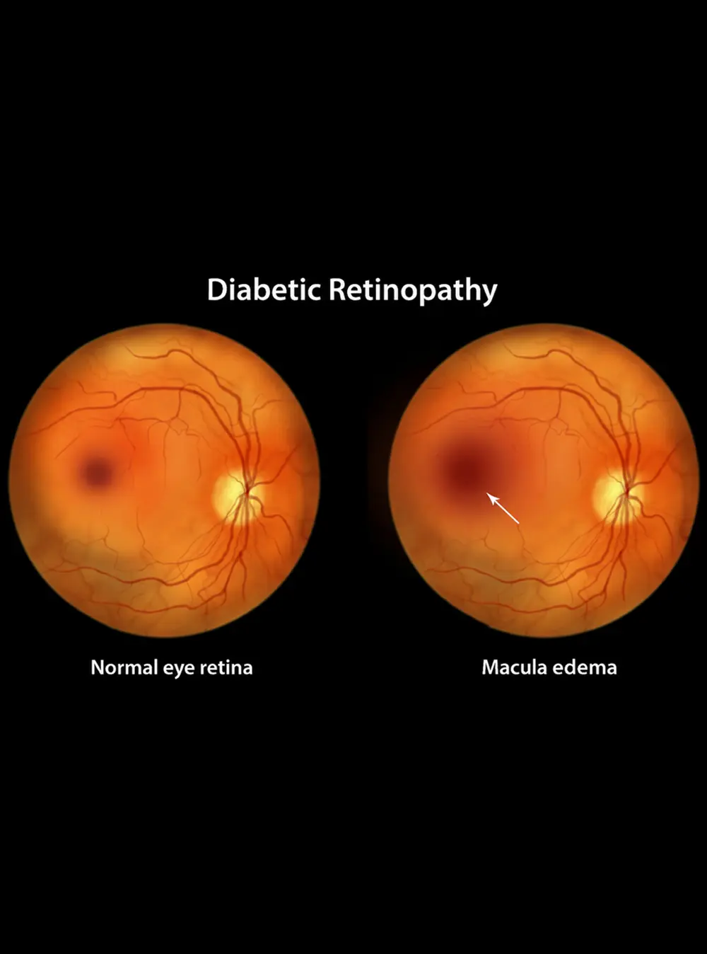
Cystoid macular edema (CME) is swelling of the macula, the central part of the retina responsible for sharp, detailed vision. The swelling occurs when fluid collects within the layers of the macula, creating small, cyst-like spaces.
Since the macula controls central vision, swelling can impair reading, face recognition, driving, and seeing fine details. Cystoid macular edema isn't a single disease; it can result from eye conditions, medical procedures, or inflammation. Identifying the root cause is essential for proper treatment.

What Causes Cystoid Macular Edema?
There are several possible causes. It can occur when changes in the retinal blood vessels allow fluid to leak into the macula.
Common causes and related conditions include:
- Eye surgery, particularly cataract surgery
- Retinal vein occlusion
- Diabetic retinopathy
- Inflammation inside the eye
- Certain retinal disorders
- Eye trauma
- Some medications
CME that develops after cataract surgery is sometimes called pseudophakic cystoid macular edema. The timing, severity, and underlying cause can vary from one patient to another. Our retina specialist can identify the cause of swelling using an exam and retinal imaging.
What Are the Symptoms of Cystoid Macular Edema?
The cystoid macular edema symptoms can range from mild blurring to noticeable changes in central vision.
You may experience:
- Blurry or hazy central vision
- Reduced visual sharpness
- Distorted or wavy vision
- Difficulty reading
- Difficulty recognizing faces
- Colors appearing less vivid
- A central area of vision that appears distorted
CME does not always cause pain. Some patients notice gradual changes, while others become aware of vision changes after an eye procedure or when an underlying retinal condition becomes active.

How Is Cystoid Macular Edema Diagnosed?
A cystoid macular edema diagnosis usually involves a comprehensive retinal examination and imaging.
Dilated Eye Examination
Your pupils are dilated so the retina and macula can be examined closely. Our specialist will look for swelling, bleeding, inflammation, or other retinal changes.
Fluorescein Angiography
In selected cases, fluorescein angiography may be performed. A dye is injected into a vein in the arm and photographed as it moves through the retinal blood vessels.
This can help identify leakage and provide additional information about the cause of the swelling.
OCT Imaging
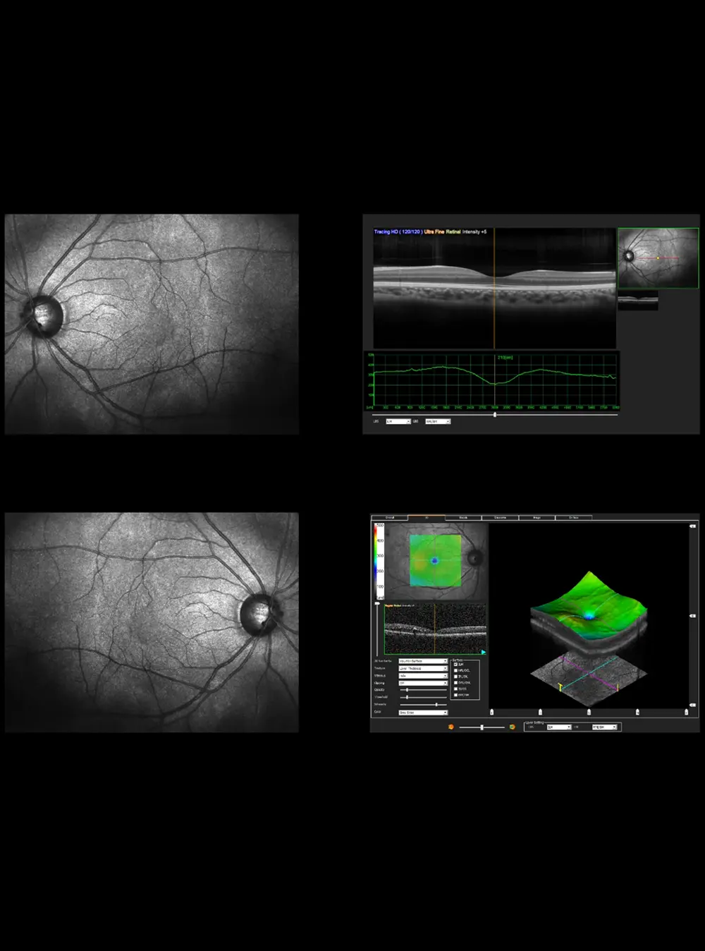
Optical coherence tomography (OCT) is a non-contact scan that creates detailed cross-sectional images of the retina.
OCT can show:
- Fluid within the macula
- The location and extent of swelling
- Changes in retinal thickness
- How the macula responds to treatment
OCT is particularly useful for monitoring CME over time.
How Is Cystoid Macular Edema Treated?
The appropriate CME treatment depends on what caused the swelling, how severe it is, and how the retina responds to treatment.
Anti-Inflammatory Treatment
When inflammation is contributing to CME, anti-inflammatory eye drops, injections, or other medications may be considered depending on the circumstances.
Retinal Injections
In some cases, medications may be injected into the eye to reduce retinal swelling and leakage. Anti-VEGF injections may be appropriate when CME is associated with conditions such as retinal vein occlusion or diabetic retinal disease. The treatment selected depends on the underlying retinal condition rather than the swelling alone.
Steroid Treatment
Corticosteroid treatment may be considered for certain patients, particularly when inflammation plays an important role. The method of treatment can vary, and our retina specialist will determine whether it is appropriate based on your examination and medical history.
Treating the Underlying Cause
Managing the condition responsible for the edema is an important part of treatment. For example, if swelling is associated with diabetic retinopathy or retinal vein occlusion, treatment may need to address both the macular swelling and the underlying retinal disease.
Monitoring
Some cases require close observation with repeat examinations and OCT imaging. Monitoring allows our specialist to determine whether the swelling is improving, remaining stable, or requiring additional treatment.
How Long Does Cystoid Macular Edema Last?
The duration of CME varies considerably.
Some cases improve within weeks or months, particularly when the underlying cause is identified and treated. Other cases may persist or recur and require longer-term management.
Your recovery depends on factors such as the cause of the edema, the severity of retinal swelling, how quickly treatment begins, and your response to treatment.
Regular follow-up helps determine whether the macula is recovering.
When Should You See a Retina Specialist?
Contact an eye doctor if you develop new or worsening changes in central vision, particularly after eye surgery or if you have an existing retinal condition.
Seek prompt evaluation if you notice:
- New blurry or distorted central vision
- Straight lines appearing wavy
- Difficulty reading
- A sudden decline in vision
- New dark or missing areas in your central vision
Do not assume that blurry vision after an eye procedure is simply part of normal recovery. Persistent or worsening symptoms should be evaluated to determine whether macular swelling or another retinal problem is present.


Why Choose Dr. Paul Hahn?
Dr. Paul Hahn, MD, PhD, FASRS, is a board-certified retina specialist with extensive experience treating retinal diseases that can affect the macula.
He is fellowship-trained in vitreoretinal surgery and has Over 9 years of surgical experience in retina care. Dr. Hahn has also led more than 30 clinical trials with over 100 publications in retinal disease treatment.
At phRETINA in Nanuet, NY, he uses advanced retinal imaging and evidence-based treatment approaches to evaluate conditions affecting the macula. His clear and respectful approach helps patients understand their diagnosis and treatment options.
Conclusion
The right treatment depends on the underlying cause and the condition of the retina. A comprehensive examination and OCT imaging can help identify the swelling and guide an appropriate treatment plan.
If you are experiencing persistent blurry or distorted vision, a retinal evaluation can help determine whether CME or another condition is responsible.
Frequently Asked Questions
Concerned About Blurry or Distorted Vision?
Schedule a comprehensive retinal evaluation today.