
Have your eyes ever had sudden floaters, cloudy vision, or a dark haze across them? If so, then it can be alarming, especially when they appear without pain.
One possible cause is vitreous hemorrhage, a condition in which blood leaks into the clear gel that fills the inside of the eye.
The important step is finding out why the bleeding occurred, because some causes require urgent treatment to protect the retina and preserve vision.

What Is Vitreous Hemorrhage?
It occurs when blood enters the vitreous, the clear, gel-like substance that fills the space between the lens and the retina.
The vitreous lets light reach the retina in the back of the eye. Blood in this area can blur or scatter light, making eyesight cloudy or patchy.
Some see dark floaters. Others lose vision suddenly and severely. The amount and location of blood will determine severity.
Although people sometimes describe this as an eye hemorrhage behind the eye, vitreous hemorrhage specifically refers to bleeding inside the vitreous cavity.
It is different from a subconjunctival hemorrhage, which causes a visible red patch on the white of the eye.
What Causes Vitreous Hemorrhage?
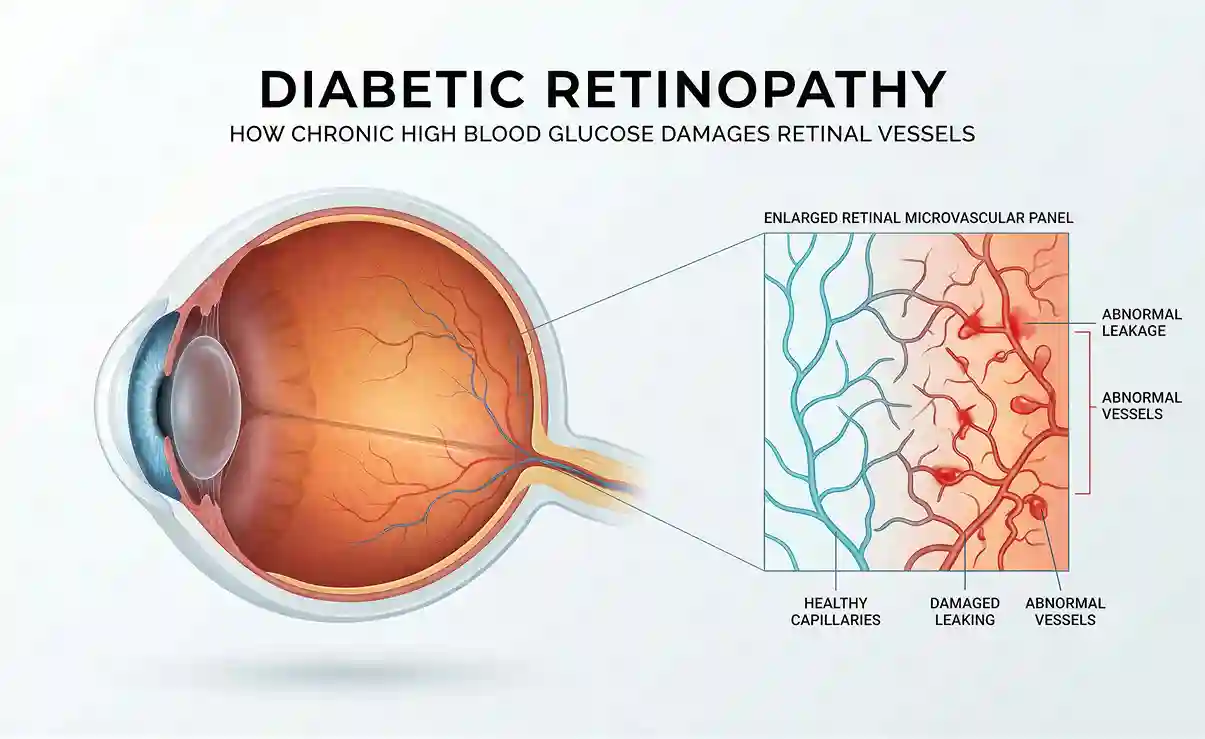
Diabetic Retinopathy
Diabetes can damage the small blood vessels that supply the retina. In advanced diabetic retinopathy, the retina may develop abnormal new blood vessels. These vessels are fragile and can bleed into the vitreous.
This is an important cause of vitreous hemorrhage, particularly in people with advanced diabetic eye disease.
Posterior Vitreous Detachment and Retinal Tears
As people age, the vitreous can gradually pull away from the retina. This process is called posterior vitreous detachment.
Sometimes, the pulling damages a retinal blood vessel or creates a retinal tear. Bleeding may occur as a result. When vitreous hemorrhage develops alongside sudden flashes or floaters, a retinal tear must be considered.
Retinal Vein Occlusion
A retinal vein occlusion occurs when a vein that carries blood away from the retina becomes blocked. This can lead to retinal bleeding, swelling, and, in some cases, the growth of fragile new blood vessels that may bleed into the vitreous.
Eye Injury
A blow to the eye or a penetrating injury can damage blood vessels and cause bleeding inside the eye. Any sudden vision change following an eye injury needs urgent assessment.
Other Retinal or Medical Conditions
Less commonly, vitreous hemorrhage may be associated with retinal inflammation, certain blood disorders, or other retinal diseases. In some cases, the bleeding may be the first sign of an underlying condition that has not yet been diagnosed.
What Are the Symptoms of Vitreous Hemorrhage?
The symptoms of vitreous hemorrhage can appear suddenly and may affect one eye. They are often painless, which can make the change in vision unexpected.
Common symptoms include:
- New floaters that look like dots, threads, cobwebs, or dark specks
- Blurred or hazy vision
- A reddish or dark tint to vision
- Shadows or areas of reduced vision
- Sudden, significant vision loss when bleeding is extensive
A vitreous hemorrhage in the left eye causes symptoms in that eye, although the underlying condition may affect both eyes over time. For example, diabetic retinopathy can involve both eyes, even if bleeding develops in only one initially.
The amount of bleeding does not always indicate how serious the underlying problem is. Even if vision loss seems mild, a retinal tear or detachment may still be present.
Is Vitreous Hemorrhage More Common in Older Adults?
Bleeding behind the eye in elderly patients can occur for several reasons. Age-related changes in the vitreous make posterior vitreous detachment more common, and this can sometimes lead to bleeding or a retinal tear.
Older adults may also have conditions such as diabetes, retinal vein occlusion, or age-related retinal disease that increase the risk of bleeding.
However, vitreous hemorrhage is not limited to older people. Younger adults can develop it after an eye injury or because of certain retinal or blood vessel conditions.
Age alone cannot identify the cause. A retinal examination is needed to determine what has happened and whether treatment is required.
What Are the Three Types of Retinal Bleeding?
These are not three formal categories of vitreous hemorrhage, but understanding the differences can help clarify the diagnosis.
- Intraretinal hemorrhage: Blood collects within the layers of the retina. It may occur with diabetic retinopathy or retinal vein occlusion.
- Subretinal hemorrhage: Blood collects beneath the retina, between the retina and the underlying tissue. It can occur with certain abnormal blood vessel conditions.
- Vitreous hemorrhage: Blood enters the clear vitreous cavity in front of the retina, where it can interfere with the passage of light and cause floaters or blurred vision.
These types of bleeding occur in different locations and may require different treatments. An eye exam can help find which condition is present.

How Is Vitreous Hemorrhage Diagnosed?
A retina specialist will ask about the onset of symptoms, previous eye conditions, diabetes, recent injuries, and other relevant medical history.
A dilated retinal examination allows the specialist to look for bleeding, retinal tears, abnormal blood vessels, and signs of retinal detachment.
If the blood blocks the view of the retina, an ultrasound scan may be needed to check whether the retina is attached. Additional retinal imaging may be performed when the view improves.
What Is the Treatment for Vitreous Hemorrhage?
The right treatment depends on the cause of the bleeding, the amount of blood, and whether the retina is damaged.
Observation and Monitoring
If the retina is attached and no urgent complication is found, a retina specialist may recommend monitoring while the blood clears naturally. The blood can take weeks or longer to clear, depending on the amount and the underlying cause.
Follow-up is essential because a retinal tear or other problem may be difficult to see while the blood obscures the view.
Laser Treatment
Laser photocoagulation may be used when abnormal retinal blood vessels or a retinal tear require treatment. For example, laser treatment can help control bleeding risk from abnormal vessels associated with diabetic retinopathy.
The laser does not directly remove blood already present in the vitreous. Instead, it treats the underlying retinal problem when appropriate.
Injections
Anti-VEGF injections may be recommended when abnormal blood vessel growth is contributing to the bleeding, such as in certain cases of diabetic retinopathy or retinal vein occlusion.
These medications help reduce abnormal vessel activity. They are not necessary for every vitreous hemorrhage, and the treatment plan depends on the underlying diagnosis.
Vitrectomy Surgery
If the blood does not clear, vision remains significantly affected, or a retinal tear or detachment requires surgery, a procedure called vitrectomy may be recommended.
During vitrectomy, the surgeon removes the blood-filled vitreous and addresses the underlying problem when possible. Additional laser treatment or other procedures may be performed during the same surgery.
The decision to operate depends on the retinal findings, the severity of vision loss, and the expected benefits and risks.
Why Choose Dr. Paul Hahn at phRETINA?
Dr. Paul Hahn, MD, PhD, FASRS, is a board-certified, fellowship-trained retina specialist with extensive experience in retinal and macular care.
He has led more than 30 clinical trials and has over 100 publications in retinal disease treatment.
At phRETINA in Nanuet, NY, Dr. Hahn combines retinal imaging with evidence-based treatment and personalized care.
The practice serves patients throughout Rockland County, Bergen County, NJ, and nearby communities in Westchester County, NY.
Conclusion
Vitreous hemorrhage can cause sudden floaters, hazy vision, or significant vision loss. It may be related to diabetes, a retinal tear, abnormal blood vessels, or an eye injury. Because some causes can threaten sight, new symptoms should never be ignored.
A retinal exam can identify the cause of the bleeding and help determine whether monitoring, injections, laser treatment, or surgery is appropriate.
FAQs
- Can vitreous hemorrhage heal on its own?
In some cases, the blood clears gradually without surgery. However, the underlying cause must be identified, and follow-up is important to check for retinal tears or detachment. - Is vitreous hemorrhage painful?
It is usually painless. Pain or other unusual symptoms should also be evaluated. - What causes leaking blood vessels in the eyes?
Fragile abnormal vessels can develop in conditions such as diabetic retinopathy and retinal vein occlusion. These vessels may leak or rupture, sometimes causing vitreous hemorrhage. - What is the treatment for a leaking blood vessel in the eye?
Treatment depends on where the bleeding occurs and what causes it. Options may include monitoring, anti-VEGF injections, laser treatment, or vitrectomy surgery. - Can vitreous hemorrhage cause permanent vision loss?
It can, particularly when the bleeding is associated with a retinal detachment or serious underlying retinal disease. Prompt diagnosis and appropriate treatment can help reduce the risk of lasting damage.