Types of Retinal Detachment
- Home
- General
Types of Retinal Detachment
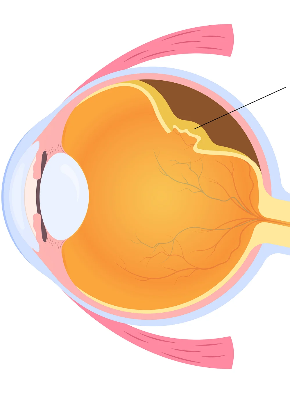
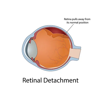
Understanding the different types of retinal detachment is important because each form develops differently and may require a different treatment approach. Retinal detachment occurs when the retina separates from the tissue beneath it, disrupting its blood supply and function.
Without prompt diagnosis and treatment, retinal detachment can lead to permanent vision loss. Recognizing the different retina detachment types helps patients understand the urgency of seeking specialized retinal care.
At phRETINA, our retina specialists diagnose and treat all forms of retinal detachment using advanced imaging and individualized treatment plans designed to preserve vision.
How Retinal Detachment Occurs
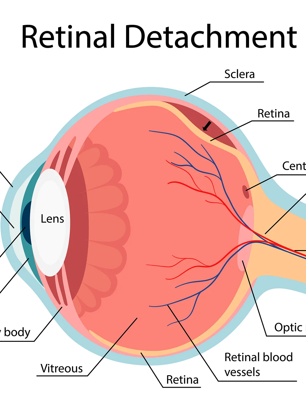
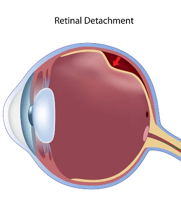
The retina is a thin layer of light-sensitive tissue located at the back of the eye. It converts light into signals that are sent to the brain, allowing us to see.
When the retina separates from the underlying tissue, it can no longer function normally. As the detachment progresses, vision may become increasingly impaired.
Although all retinal detachments involve separation of the retina, the underlying causes differ significantly. This is why understanding the different types of retinal detachment is essential for both diagnosis and treatment planning.


The Three Main Types of Retinal Detachment
The primary retina detachment types include three distinct forms, each developing through a different mechanism and presenting unique clinical challenges.
The Three Types
- Rhegmatogenous retinal detachment
- Tractional retinal detachment
- Exudative retinal detachment
Accurate diagnosis is essential because treatment strategies differ depending on the specific type of retinal detachment.
Rhegmatogenous Retinal Detachment
Rhegmatogenous retinal detachment is the most common form of retinal detachment and develops when a retinal tear or hole allows fluid to collect beneath the retina.
Common Causes
- Age-related vitreous changes
- Severe nearsightedness
- Previous eye surgery
- Eye trauma
- Prior retinal tears
Symptoms
- Sudden floaters
- Flashes of light
- Blurred vision
- A curtain or shadow across vision


Tractional Retinal Detachment
Tractional retinal detachment occurs when scar tissue on the retinal surface pulls the retina away from the back of the eye. Unlike rhegmatogenous retinal detachment, this condition often develops gradually.
Common Causes
- Diabetic retinopathy
- Retinal vascular disease
- Previous retinal surgery
- Inflammatory eye conditions
Symptoms
- Gradual vision loss
- Distorted vision
- Reduced peripheral vision
- Progressive visual changes
Patients with advanced diabetic eye disease are among those at highest risk for developing tractional retinal detachment.

Exudative Retinal Detachment
Exudative retinal detachment develops when fluid accumulates beneath the retina without a retinal tear being present.
How It Develops
Inflammation, tumors, vascular abnormalities, or other underlying medical conditions cause fluid to leak beneath the retina.
Common Causes
- Inflammatory diseases
- Retinal vascular disorders
- Tumors
- Central serous chorioretinopathy
- Severe hypertension
Symptoms
- Blurred vision
- Distorted vision
- Visual field defects
- Changes in central vision
Because no retinal tear is present, treatment focuses on addressing the underlying condition responsible for the fluid accumulation.
Comparing the Types of Retinal Detachment
While all types of retinal detachment threaten vision, their causes, symptoms, and treatment strategies differ significantly.
Rhegmatogenous Retinal Detachment
- Caused by a retinal tear or hole
- Most common type
- Often associated with flashes and floaters
Tractional Retinal Detachment
- Caused by scar tissue pulling on the retina
- Frequently associated with diabetic eye disease
- Usually develops gradually
Exudative Retinal Detachment
- Caused by fluid accumulation beneath the retina
- No retinal tear present
- Often linked to underlying medical conditions
Accurate diagnosis is essential because each type requires a different treatment approach.


Warning Signs of Retinal Detachment
Regardless of the type of retinal detachment, many patients experience similar warning signs that require immediate medical attention.
Common Symptoms Include
- Sudden floaters
- Flashes of light
- Blurred vision
- Distorted vision
- Loss of peripheral vision
- A curtain-like shadow across vision
These symptoms may indicate a detached retina and should always be evaluated promptly by a retina specialist.
How Retinal Detachment Is Diagnosed
Prompt diagnosis is critical whenever retinal detachment is suspected. Early evaluation allows treatment to begin before permanent vision loss occurs.
Diagnostic Evaluation May Include
- Comprehensive dilated retinal examination
- Optical Coherence Tomography (OCT)
- Wide-field retinal imaging
- Ocular ultrasound
- Assessment of retinal tears and retinal health
At phRETINA, advanced diagnostic technology enables our specialists to accurately identify the specific type of retinal detachment and determine the most appropriate treatment plan.


Treatment for Retinal Detachment
Treatment depends on the specific type of retinal detachment and the severity of the condition. The primary goal is to preserve vision, repair retinal damage, and reduce the risk of recurrent detachment.
Laser & Cryotherapy
- Laser treatment for retinal tears
- Cryotherapy
Retinal Surgery
- Pneumatic retinopexy
- Vitrectomy surgery
- Scleral buckle surgery
Medical Management
- Treatment of inflammatory disease
- Management of retinal vascular disease
- Long-term retinal monitoring
At phRETINA, treatment recommendations are individualized based on the patient's retinal condition, visual symptoms, and long-term goals for preserving vision.


Why Early Detection Matters
Early diagnosis plays a critical role in preserving vision and improving treatment outcomes. Seeking prompt care when symptoms develop can significantly increase the chances of successful retinal repair.
Benefits of Early Detection
- Greater likelihood of successful retinal repair
- Reduced risk of permanent vision loss
- Earlier treatment of retinal tears
- Improved long-term visual outcomes
- Prevention of disease progression
The sooner retinal detachment is diagnosed and treated, the better the opportunity to preserve vision and reduce the risk of long-term complications.
When to Seek Evaluation
You should schedule an urgent retinal evaluation if you experience any symptoms that may indicate a retinal tear or retinal detachment.
Seek Immediate Care If You Notice:
- New floaters
- Flashes of light
- Sudden blurred vision
- Peripheral vision loss
- A shadow or curtain across vision
- Symptoms of a retinal tear or retinal detachment
Because retinal detachment is a medical emergency, prompt evaluation by a retina specialist is essential to protect your vision.


Helpful Information for New and Returning Patients
Schedule a Retinal Evaluation
If you are experiencing symptoms of retinal detachment or have concerns about a retinal tear, a comprehensive retinal evaluation can help determine the cause and guide the most appropriate treatment.