
Laser Retinopexy for Retinal Tears & Holes
- Home
- General

Laser Retinopexy for Retinal Tears and Holes
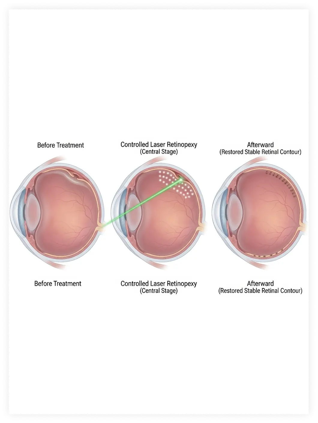
A retinal tear or hole can create a pathway through which fluid may move underneath the retina, potentially leading to a retinal detachment. Laser retinopexy is a retinal laser treatment that creates a series of small laser burns around a retinal tear or hole. These burns form a barrier of scar tissue that helps secure the retina and reduce the risk of progression to retinal detachment.
Laser retinopexy is commonly performed in an ophthalmologist's office and does not require an incision. The treatment recommended depends on the location and characteristics of the retinal break and the overall condition of the retina.
What Is Laser Retinopexy?
Laser retinopexy is a procedure used to treat certain retinal tears and holes before they progress to retinal detachment. During the laser retinopexy procedure, focused laser light is applied around the retinal break. The laser produces controlled burns that eventually develop into scar tissue.
This creates an adhesion between the retina and the underlying tissue, helping prevent fluid from passing through the tear and separating the retina. The procedure is different from retinal detachment surgery.
When Is Laser Retinopexy Needed?
Laser retinopexy may be recommended when an examination identifies a retinal tear or hole that presents a meaningful risk of retinal detachment.
Laser Retinopexy for Retinal Tear
A retinal tear can occur when the vitreous, the gel that fills the inside of the eye, pulls on the retina. Some retinal tears can progress to retinal detachment if fluid passes through the opening. Laser retinopexy for retinal tear seals the area around the break to help reduce this risk. Not every retinal tear requires laser treatment. The decision depends on factors such as the appearance and location of the tear, symptoms, surrounding retinal changes, and individual risk factors.
Laser Retinopexy for Retinal Hole
A retinal hole is a full-thickness opening in the retina that may develop through different mechanisms. Some holes have a low risk of causing retinal detachment, while others may warrant treatment. Laser retinopexy for retinal hole can be used to create a protective barrier around a hole when treatment is considered appropriate.
Laser Retinopexy for Lattice Degeneration
Lattice degeneration is a thinning pattern that develops in areas of the peripheral retina. It does not automatically require treatment. In selected patients, laser retinopexy for lattice degeneration may be considered when there are associated retinal breaks or other features that increase concern for retinal detachment. our retina specialist will determine whether observation or treatment is appropriate.

Retinal Tear and Retinal Hole Symptoms
Some retinal tears and holes cause no symptoms and are discovered during a dilated eye examination. Others can produce noticeable changes in vision.
Possible retinal hole symptoms and symptoms associated with retinal tears include:
- A sudden increase in floaters
- Flashes of light
- New spots or shadows in the vision
- A curtain or veil moving across the field of vision
- Sudden changes in peripheral vision
A sudden increase in floaters or flashes, especially when accompanied by a shadow or curtain in the vision, requires urgent retinal evaluation.
What Happens During the Laser Retinopexy Procedure?
Before treatment, the eye is examined carefully to identify the location and extent of the retinal break.
During retinal laser treatment:
- 1. The eye is dilated so the retina can be examined and treated.
- 2. Numbing drops are used to reduce discomfort.
- 3. A specialized lens may be placed on the eye to help the physician view the retina.
- 4. Focused laser applications are placed around the retinal tear or hole.
- 5. The treatment creates a pattern of controlled burns surrounding the affected area.
The procedure generally does not involve cutting into the eye.

Does Laser Retinopexy Hurt?
Patients may experience some pressure or discomfort during treatment, but laser retinopexy pain is generally limited because numbing medication is used.
You may notice bright flashes of light while the laser is being applied. Some patients describe temporary aching or irritation afterward. If you experience significant or worsening pain after treatment, contact the treating eye specialist promptly.
Laser Retinopexy Recovery
Laser retinopexy recovery is usually relatively short because the procedure does not require an incision.
However, the laser scars need time to develop and strengthen. For this reason, patients may be advised to avoid certain strenuous activities for a period determined by their physician. Vision can also be temporarily different following treatment.

Blurry Vision After Laser Retinopexy
Blurry vision after laser retinopexy can occur temporarily, particularly because the pupil is dilated and the eye may be irritated following treatment.
Some patients may notice temporary floaters, light sensitivity, or visual changes. These symptoms should gradually improve.
New or worsening vision loss, a sudden increase in floaters or flashes, or a curtain-like shadow is not something to simply attribute to recovery. These symptoms require prompt evaluation.
Laser Retinopexy Aftercare
Laser retinopexy aftercare instructions vary according to the location of the retinal tear or hole and the patient's individual circumstances.
After treatment, our physician may recommend:
- Following activity restrictions for the recommended period
- Using prescribed eye drops when indicated
- Avoiding rubbing the treated eye
- Attending scheduled follow-up examinations
- Monitoring for new flashes, floaters, shadows, or vision loss
Follow-up is important because laser treatment does not prevent new retinal tears from developing elsewhere in the eye.


Laser Retinopexy Risks
Laser retinopexy is generally considered a well-established treatment for appropriate retinal breaks, but like any medical procedure, it has potential risks.
Possible laser retinopexy risks include:
- Temporary blurred vision
- Temporary eye discomfort
- Inflammation
- Increased eye pressure
- Visual symptoms related to the laser treatment
- Incomplete treatment of the retinal break
- Development of additional retinal tears
- Retinal detachment despite treatment
The specific risks depend on the condition being treated and the location of the retinal break.
Laser Retinopexy vs. Retinal Detachment Surgery
Laser retinopexy is generally used to treat certain retinal tears or holes before a retinal detachment develops.
If the retina has already detached, laser alone may not be sufficient. Treatment may involve procedures such as pneumatic retinopexy, scleral buckle surgery, or vitrectomy, depending on the type and extent of detachment. Early evaluation of retinal tear symptoms is therefore important.
When Should You See a Retina Specialist?
Seek urgent eye care if you experience:
Warning Signs
- Sudden flashes of light
- A sudden increase in floaters
- New peripheral vision loss
Vision Changes
- A dark curtain or shadow across your vision
- Sudden or unexplained vision loss
These symptoms can indicate a retinal tear or retinal detachment, both of which require prompt evaluation.
If you are searching for retinal laser treatment, retinal hole repair, or laser retinopexy near me, a comprehensive retinal examination can determine whether laser treatment is appropriate.
Why Choose Dr. Paul Hahn?
Dr. Paul Hahn, MD, PhD, FASRS, is a board-certified retina specialist with extensive experience treating retinal conditions. He is fellowship-trained in vitreoretinal surgery and has led more than 30 clinical trials with over 100 publications in retinal disease treatment.
At phRETINA in Nanuet, NY, he combines advanced retinal imaging with evidence-based treatment and individualized patient care.
The approach focuses on identifying retinal problems early and explaining treatment options clearly so patients can make informed decisions about their eye health.
Conclusion
Laser retinopexy for retinal tear is a commonly used retinal laser treatment designed to create a protective barrier around certain retinal tears and holes. By treating the retina before a detachment occurs, the procedure can help reduce the risk of retinal detachment in appropriately selected patients.
Because retinal tears and holes can sometimes occur without obvious symptoms, regular dilated retinal examinations are important for patients at increased risk. Sudden flashes, new floaters, shadows, or a curtain over the vision should always be evaluated urgently.