
Diabetic retinopathy is one of the leading causes of vision loss among adults with diabetes. The challenge is that the condition often develops silently, causing little to no noticeable symptoms until significant retinal damage has already occurred. This is why retinal imaging has become an essential part of modern diabetic eye care.
Advanced retinal imaging allows our retina specialist to detect the earliest signs of Diabetic retinopathy often before vision is affected.
By identifying subtle changes in the retina, treatment can begin sooner, helping reduce the risk of permanent vision loss.

What Is Retinal Imaging?
Retinal imaging refers to a group of advanced diagnostic tests that capture detailed images of the retina, the light-sensitive tissue at the back of the eye.
Unlike a standard vision test, retinal imaging allows our retina specialist to examine the retina in remarkable detail, identifying changes that may not yet be causing symptoms. For people living with diabetes, these images play an important role in detecting diabetic retinopathy early and monitoring changes over time.
Why Early Detection Matters
Diabetic retinopathy occurs when high blood sugar damages the tiny blood vessels in the retina. Over time, these blood vessels can leak fluid or blood, reducing the retina's ability to function properly.
In the early stages, many people experience no noticeable diabetic retinopathy symptoms. As the disease progresses, patients may develop blurred vision, floaters, dark spots, or vision loss.
Because symptoms often appear after retinal damage has already occurred, regular retinal imaging is one of the most effective ways to detect diabetic retinopathy before it affects your sight.

How Retinal Imaging Detects Diabetic Retinopathy
Modern retinal imaging provides detailed information about the health of your retina and helps identify early signs of diabetic eye disease.
Fundus Photography
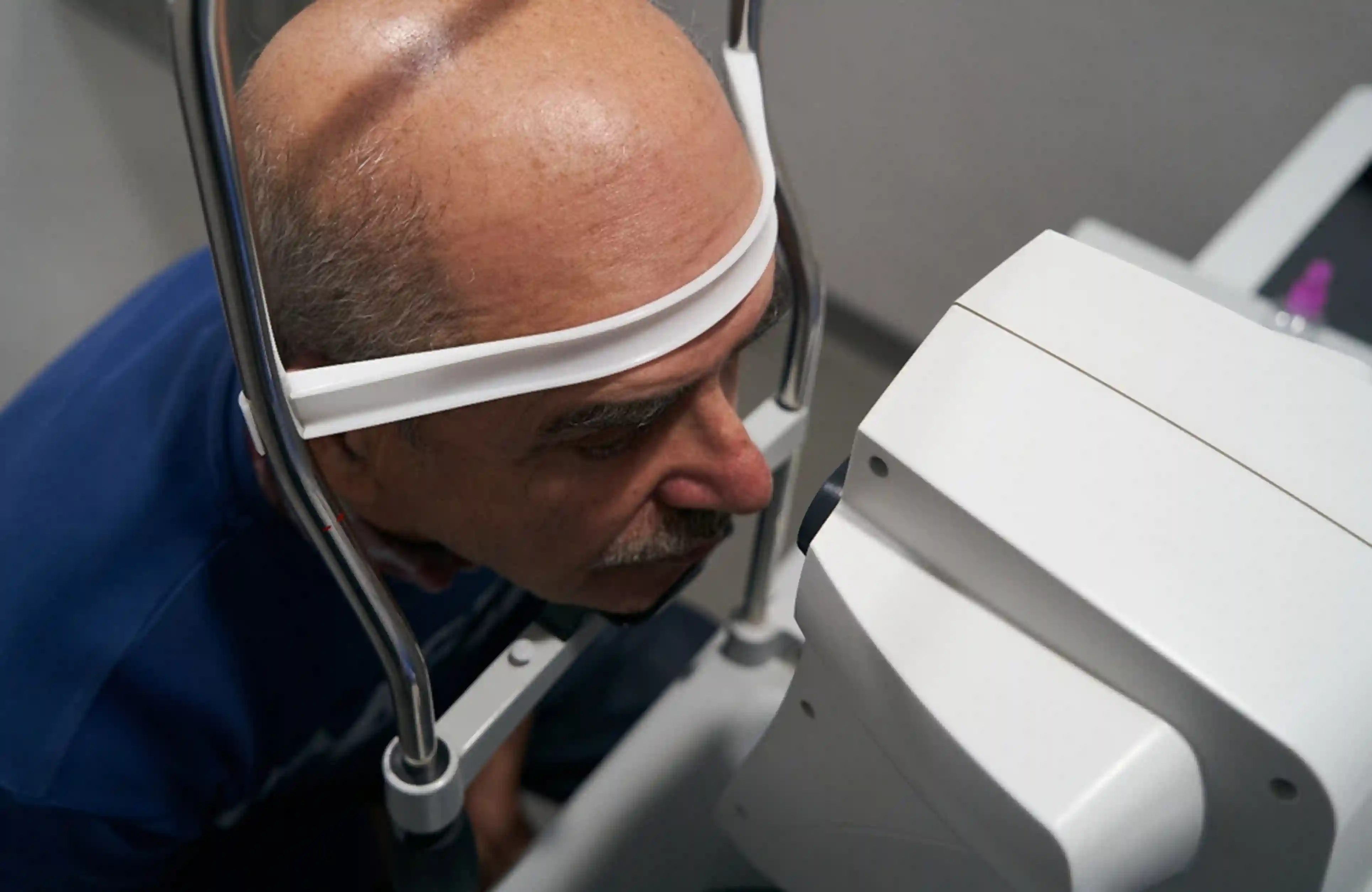
Fundus photography captures high-resolution color images of the retina. These photographs allow our retina specialist to detect small hemorrhages, leaking blood vessels, swelling, and other early signs of diabetic retinopathy.
By comparing images over time, fundus photography also helps monitor disease progression and evaluate how well treatment is working.
OCT Eye Scan
An OCT eye scan creates detailed cross-sectional images of the retina using light waves.
This technology allows our retina specialist to identify retinal swelling, fluid buildup, and structural changes that may not be visible during a routine examination. OCT is especially valuable for detecting diabetic macular edema, a common complication of diabetic retinopathy that can affect central vision.
Dilated Retinal Exam
A dilated retinal exam remains an essential part of evaluating diabetic eye disease.
After your pupils are dilated with eye drops, our retina specialist can carefully examine the retina, optic nerve, and retinal blood vessels for signs of bleeding, swelling, or abnormal blood vessel growth.
Retinal imaging and a dilated retinal exam work together to provide a comprehensive evaluation of retinal health.
The Role of a Comprehensive Eye Examination
Although retinal imaging is highly effective, it should be part of a comprehensive eye examination rather than a replacement for one.
A complete diabetic eye evaluation may include:
- A review of your medical and vision history
- Visual acuity testing
- Retinal imagingv
- Fundus photography
- OCT eye scan
- Dilated retinal exam
- Assessment of retinal blood vessels
Together, these tests provide a detailed understanding of your eye health and help detect changes before they threaten your vision.
Who Should Have Retinal Imaging?
Anyone with diabetes should schedule regular eye examinations, even if their vision seems normal.
Retinal imaging is particularly important for:
- People with Type 1 diabetes
- People with Type 2 diabetes
- Individuals with gestational diabetes
- Patients with existing diabetic retinopathy
- Anyone experiencing changes in vision
Regular imaging allows our retina specialist to monitor for retinal damage and recommend treatment if changes are detected early.
Benefits of Retinal Imaging for People with Diabetes
Regular retinal imaging offers significant benefits because it helps detect changes in the retina before they affect your vision.
Some of the key advantages include:
- Detecting diabetic retinopathy in its earliest stages
- Identifying retinal damage before symptoms develop
- Monitoring changes over time
- Guiding personalized treatment decisions
- Helping reduce the risk of permanent vision loss
Since diabetic eye disease can progress without noticeable symptoms, routine retinal imaging allows our retina specialist to monitor your retinal health and recommend treatment at the most appropriate time.
When Should You See a Diabetic Retina Specialist?
If you have diabetes, routine eye care is essential, even if your vision seems normal. A regular diabetic eye exam combined with advanced retinal imaging can help identify retinal changes before they become more serious.
You should schedule an evaluation if you:
- Have Type 1 or Type 2 diabetes
- Have been diagnosed with diabetic retinopathy
- Notice blurred vision or new floaters
- Experience difficulty reading or focusing reading or focusing
- Have not had a dilated retinal exam within the past year
If diabetic retinopathy is suspected, referral to a diabetic retina specialist can help ensure you receive specialized evaluation and treatment.
At phRETINA, Dr. Paul Hahn uses advanced retinal imaging to diagnose and manage diabetic retinal disease, helping patients in Nanuet, NY, protect their vision through personalized, evidence-based care.
Conclusion
Because diabetic retinopathy often develops without noticeable symptoms, regular retinal imaging plays a critical role in protecting your vision. Technologies such as fundus photography, an OCT eye scan, and a dilated retinal exam allow our retina specialist to detect retinal damage early, monitor disease progression, and recommend treatment before permanent vision loss occurs.
If you have diabetes, scheduling routine eye examinations is one of the most effective steps you can take to preserve your long-term vision. Early detection and ongoing monitoring can make a significant difference in maintaining healthy eyesight.
Frequently Asked Questions
-
What is retinal imaging?
Retinal imaging uses specialized technology to capture detailed images of the retina. These images help detect diabetic retinopathy, retinal damage, and other retinal conditions before they cause noticeable vision changes. -
Can retinal imaging detect diabetic retinopathy early?
Yes. One of the greatest benefits of retinal imaging is its ability to detect diabetic retinopathy before symptoms develop. Early detection allows treatment to begin sooner and helps reduce the risk of vision loss. -
Is retinal imaging part of a diabetic eye exam?
Yes. A comprehensive diabetic eye exam may include retinal imaging, a dilated retinal exam, fundus photography, and an OCT eye scan to evaluate the health of the retina. -
How often should people with diabetes have retinal imaging?
Most people with diabetes should have a comprehensive eye examination at least once a year. Our retina specialist may recommend more frequent retinal imaging depending on your eye health and the severity of diabetic retinopathy. -
What are the symptoms of diabetic retinopathy?
Early diabetic retinopathy symptoms may not be noticeable. As the condition progresses, symptoms can include blurred vision, floaters, dark spots, reduced night vision, and vision loss. Regular retinal imaging can detect changes before these symptoms appear.