
When retinal problems affect vision, removing the eye's vitreous gel can sometimes help the retina heal. Vitrectomy surgery is used to treat conditions such as retinal bleeding, scar tissue, and retinal detachment.
some patients get questions in their mind when they hear about surgery. What happens during the procedure? Will vision improve right away? How long does recovery take? Is it safe?
Learn about vitrectomy and its process in this blog.
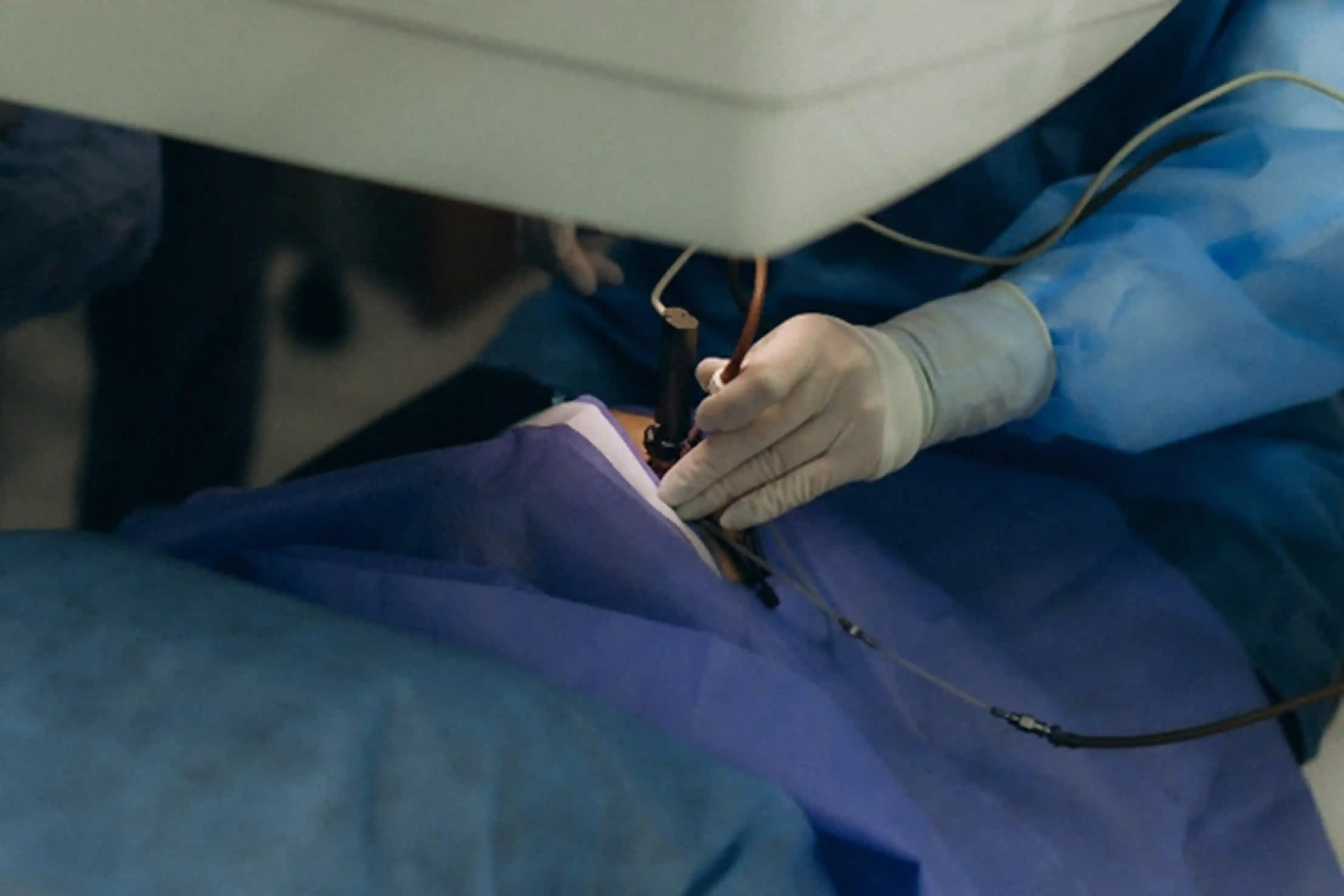
What is a Vitrectomy?
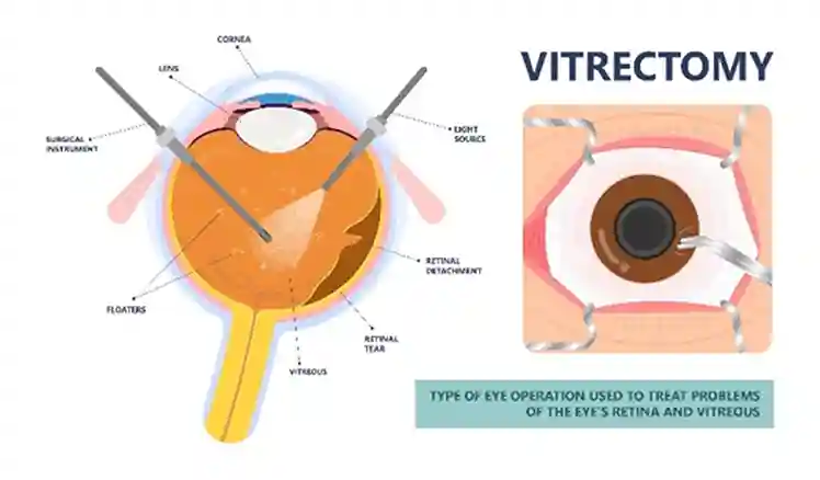
A vitrectomy is a surgical procedure in which the vitreous gel is removed from inside the eye.
The vitreous normally fills the space between the lens and retina. Although it is clear and usually does not interfere with vision, problems can grow within it. Blood may collect in the vitreous, scar tissue may form, or the vitreous may pull on the retina.
The surgeon is able to operate simply on the retina by removing the vitreous.
Depending on the reason for surgery, the procedure may also involve:
- Repairing a retinal tear or retinal detachment
- Removing scar tissue or a membrane
- Treating retinal bleeding
- Applying laser treatment
- Placing a gas bubble
A vitrectomy is not a treatment for one specific eye condition. It is a surgical technique used for several different retinal problems.
When is Vitrectomy Surgery Needed?
The signs for vitrectomy depend on what is happening inside the eye and how much the retina or vision is being affected.
Some common conditions treated with vitrectomy include:
Retinal Detachment
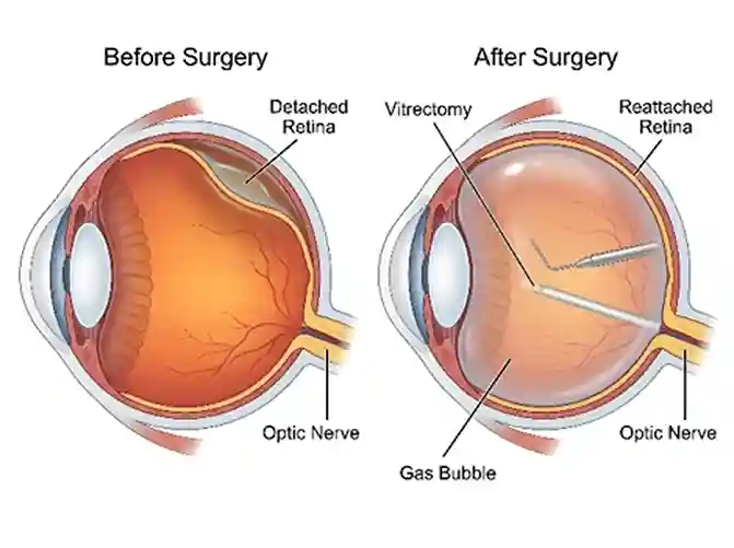
A retinal detachment occurs when the retina separates from the tissue underneath it. In certain cases, vitrectomy can remove the vitreous traction leading to the detachment and allow the surgeon to repair retinal tears and reattach the retina.
A gas bubble or silicone oil may be used to help keep the retina in position while it heals.
Vitreous Hemorrhage
A vitreous hemorrhage occurs when blood enters the vitreous. It can cause sudden or significant vision changes.
If the blood does not clear well, or is connected with another retinal problem, then vitrectomy can be performed to remove the blood and treat its underlying cause.
Macular Hole
A macular hole is a small opening in the center of the retina responsible for detailed vision. It can cause blurred or distorted central vision.
During surgery, the vitreous may be removed and a membrane on the surface of the retina may be peeled. A gas bubble may then be placed to support the macula as it heals.
Epiretinal Membrane
An epiretinal membrane is a thin layer of scar-like tissue that can form over the macula. It may cause straight lines to appear bent or make detailed vision less clear.
Vitrectomy can provide access to the membrane so it can be carefully removed when surgery is appropriate.
Advanced Diabetic Retinopathy
Diabetic retinopathy can sometimes cause significant bleeding or scar tissue inside the eye. In more advanced cases, vitrectomy may be needed to clear blood or relieve traction on the retina.
What is Vitrectomy Recovery Like?
Vitrectomy recovery varies considerably. The reason for surgery, the condition of the retina, and whether a gas bubble or silicone oil was used can all affect the recovery period.
It is normal for vision to be blurry immediately after vitrectomy eye surgery. The eye may also feel irritated or uncomfortable, and some redness or swelling can occur.
Vision does not always improve immediately. In many cases, the retina needs time to heal, and visual improvement may continue over several weeks or months.
During recovery, our retina specialist may recommend:
- Using prescribed eye drops
- Attending follow-up appointments
- Avoiding heavy lifting or strenuous activity temporarily
- Following specific head-positioning instructions when required
- Avoiding driving until vision has recovered sufficiently
Recovery instructions are individualized because different retinal surgeries require different precautions.
What if a Gas Bubble is Used?
A gas bubble is sometimes placed inside the eye after vitrectomy surgery to support the retina while it heals.
If a gas bubble is present, vision may stay blurred until the bubble slowly goes away. The amount of time this takes depends on the type of gas used.
There is one important precaution: Avoid air travel and higher altitudes while the gas bubble is in your eye.
Changes in air pressure can cause the bubble to expand and dangerously increase pressure inside the eye.
Is Vitrectomy Safe?
A common question is, “Is vitrectomy safe?”
Vitrectomy is an established retinal surgery, but no surgical procedure is completely free of risk. The potential risks also depend on why the surgery is being performed and the condition of the eye.
Possible complications include:
- Infection
- Bleeding
- Increased eye pressure
- Cataract development or progression
- Retinal tear or detachment
- Inflammation
- Macular swelling
- Low eye pressure
- Need for additional surgery
The potential risks of surgery need to be considered alongside the risks of leaving the underlying retinal condition untreated.
Our retina specialist can explain what the procedure involves, why it is being recommended, and what risks are relevant to the individual case.
Does Vitrectomy Improve Vision?
The goal of vitrectomy is to treat the underlying retinal problem and, when possible, protect or improve vision.
However, the amount of visual improvement varies. It depends on the condition being treated, how much damage has already occurred, and the health of the retina before surgery.
For example, removing blood from the vitreous may improve the view through the eye, while surgery for a macular hole is intended to allow the central retina to heal. In other situations, the main goal may be to prevent further retinal damage.
This is why visual recovery should be discussed in the context of the specific retinal condition rather than surgery alone.
When is Vitrectomy Urgent?
The urgency depends on the condition requiring surgery.
A retinal detachment, for example, requires quick treatment because delaying treatment can increase the risk of permanent vision loss.
Seek urgent retinal checkup if you experience:
- Sudden flashes of light
- A sudden increase in floaters
- A dark curtain or shadow across your vision
- Sudden peripheral vision loss
- Sudden or significant vision loss
These symptoms can occur with retinal tears or retinal detachment and should not be ignored.
Why Choose phRETINA and Dr. Paul Hahn?
Dr. Paul Hahn, MD, PhD, FASRS, is a board-certified, fellowship-trained retina specialist with extensive experience in retinal and macular care.
He has led more than 30 clinical trials and has over 100 publications in retinal disease treatment.
At phRETINA in Nanuet, NY, Dr. Hahn combines retinal imaging with evidence-based treatment and personalized care. The practice serves patients throughout Rockland County, Bergen County, NJ, and nearby communities in Westchester County, NY.
Conclusion
Vitrectomy surgery can play an important role in treating several serious retinal conditions, including retinal detachment, vitreous hemorrhage, macular hole, epiretinal membrane, and advanced diabetic retinopathy.
The vitrectomy procedure is removing the vitreous so the surgeon can access and treat the retina. Recovery changes from one patient to another, and vision may take weeks or months to improve based on the condition.
If vitrectomy has been recommended. A detailed retinal evaluation can help determine the most appropriate treatment for the condition.
FAQs
- What is a vitrectomy?
A vitrectomy is an eye surgery that removes the vitreous gel from inside the eye so the surgeon can access and treat the retina. - What conditions can vitrectomy surgery treat?
Vitrectomy may be used to treat retinal detachment, vitreous hemorrhage, macular hole, epiretinal membrane, and certain advanced complications of diabetic retinopathy. - Is vitrectomy surgery painful?
It is performed with anesthesia, and Dr. Paul Hahn uses treatment approaches designed to make the procedure as comfortable as possible. The procedure is usually painless, although some patients may experience mild irritation, soreness, or a sensation during the early recovery period. - How long does vitrectomy recovery take?
Recovery depends on the condition being treated and the type of surgery performed. Some aspects of recovery take several weeks, while visual improvement can continue for months. - Can vision be blurry after vitrectomy?
Yes. Blurry vision is common immediately after surgery, particularly when a gas bubble is used. Vision generally changes as the eye heals and the bubble gradually disappears.