Macular hole stages describe how a small structural defect in the center of the retina progresses over time. A macular hole in eye affects the macula, the part responsible for sharp central vision. It does not cause pain, but it does distort sight in ways that are hard to ignore once they begin.
Many patients first notice blurry or distorted central vision. Straight lines may appear bent. Reading becomes frustrating. These early changes are subtle, which is why timely vision eye care is important. Understanding the stages of macular hole helps patients recognize when action is needed.
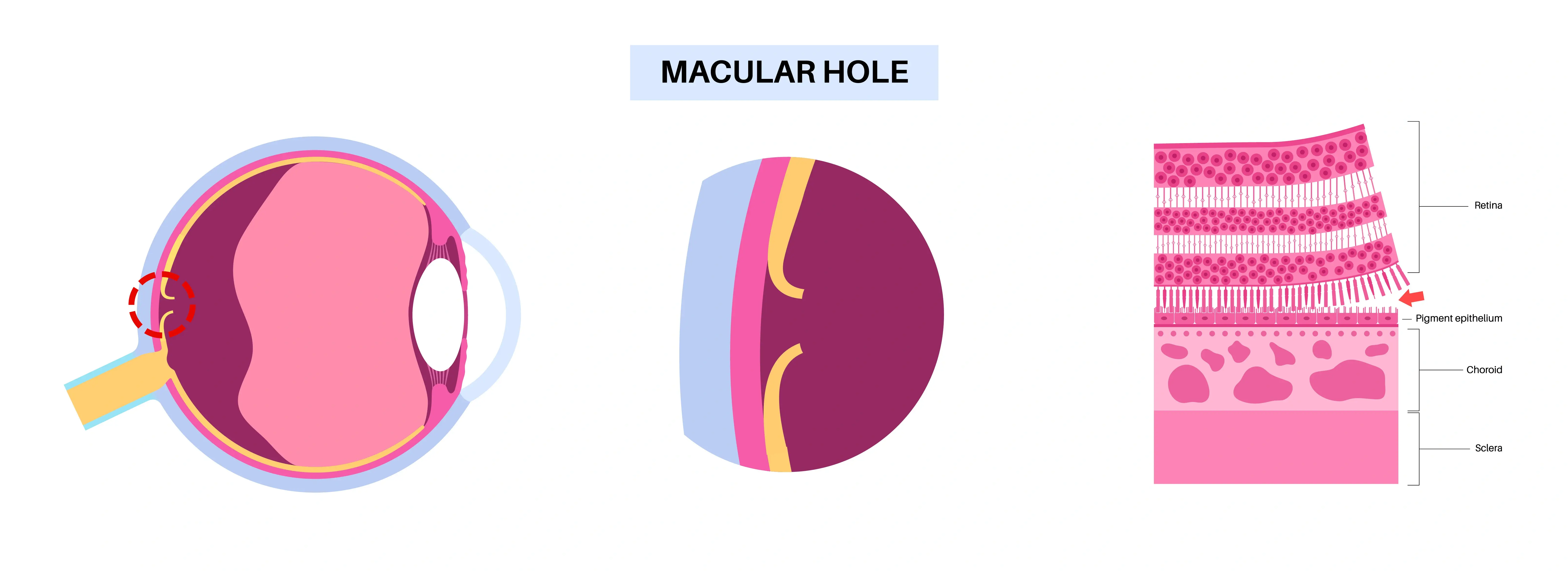
What Is a Macular Hole?
A macular hole is a small opening that forms in the macula due to traction from the vitreous gel inside the eye. As we age, the vitreous shrinks and pulls away from the retina. In some cases, the pulling force is strong enough to create an eye hole in the macular region.
Symptoms often begin gradually. People may experience blurry central vision, mild distortion, or difficulty focusing on fine detail. Some may notice floaters in vision, which can signal vitreous changes occurring at the same time. Floaters alone do not mean a macular hole, but new visual disturbances should always be evaluated.
The Four Macular Hole Stages
Macular hole stages are classified from Stage 1 to Stage 4. Each stage reflects increasing severity.
-
Stage 1: Impending Macular Hole
Stage 1 is not a full hole yet. The vitreous is pulling on the macula, causing a small central change. The retinal surface remains mostly intact. -
Stage 2: Small Full Thickness Hole
In Stage 2, a small, full thickness macular hole in eye has formed. The defect typically measures less than 400 microns. -
Stage 3: Enlarging Macular Hole
Stage 3 represents a larger full thickness hole, typically greater than 400 microns. The vitreous may still be partially attached. -
Stage 4: Advanced Macular Hole
In Stage 4, the vitreous has fully separated from the retina, and the macular hole remains open.
Symptoms at Different Stages
- Slight blurry vision
- Mild to pronounced distortion
- Central dark spot in advanced stages
- Difficulty reading or focusing on detail
What Causes a Macular Hole?
Age related vitreous changes are the most common cause. As the vitreous gel shrinks, uneven traction can create stress on the macula. Other contributing factors include high myopia, trauma, previous eye surgery, and certain retinal eye issues.
Most cases occur in individuals over 60. Women are slightly more affected than men. The process is mechanical, not inflammatory. The retina is being pulled, and at some point, tissue gives way.
Macular Hole Treatment
The primary treatment for advanced macular hole stages is vitrectomy surgery. During this procedure, a retina specialist removes the vitreous gel to eliminate traction. A gas bubble is then placed inside the eye to help the macula close properly.
After surgery, patients often traditionally have been asked need to maintain a face down position for several days. This positioning allows the gas bubble to press against the macular hole and promote healing. Dr Hahn employs advanced techniques that may minimize or eliminate the need to maintain face down positioning after surgery.
Success rates are high, particularly when surgery is performed early. Smaller holes and shorter symptom duration typically lead to better visual outcomes.
Recovery and Visual Improvement
Vision does not return instantly after macular hole treatment. Improvement happens gradually over weeks or months. Some distortion may persist, especially in later stages. However, many patients regain functional reading vision.
Regular follow up with a retina specialist ensures proper healing and monitors for additional retinal concerns. Even after closure, ongoing vision eye care remains important.
When to Seek Evaluation
Any sudden central blurry or distorted vision should not be ignored. Unlike many eye issues, the macular hole in eye affects central detail while peripheral vision often remains intact. That pattern is a key clue.
Early detection during the initial macular hole stages significantly improves the likelihood of successful treatment. Waiting allows progression, and later stages become more challenging to manage.
The retina does not send pain signals. It signals through visual distortion. Paying attention to those changes and seeking prompt care can preserve central sight and prevent long term complications.